STEMCELL Technologies STEMdiff STEMdiff Choroid Plexus Organoid Differentiation Kit
- 研究用
STEMdiff™ Choroid Plexus Organoid Differentiation Kit(製品コード:ST-100-0824)は、血液-脳脊髄液関門を形成する脳の特殊上皮「脈絡叢」にパターン化された、ヒト多能性幹細胞(hPSC)由来のオルガノイドを作製する無血清培地のキットです。
脈絡叢オルガノイドを使用すると、ヒト神経バイオマーカーの発見や中枢神経系(CNS)の透過性に関して、in vitroでアプローチ可能になります。3次元組織構造と機能的なバリア上皮を備えたオルガノイドをもちいることで、薬剤分布研究におけるCNS透過性解析や、脳脊髄液(CSF)の産生・抽出において、これまでにないアクセスと制御を実現できます。
本品の簡易な5段階のプロトコルでは、さまざまな下流用途向けの脈絡叢オルガノイドを再現性よく効率的に作製できます。
本品は、実験動物の削減、代替、改良における画期的な成果として3R賞を受賞した、Pellegrini et al.(Science, 2020)の組成に基づいています。
脈絡叢オルガノイドの形成は、中間的な胚様体(EB)の形成から開始され、神経上皮の増殖、脈絡叢様上皮へのパターン化に続きます。成熟期間の後、オルガノイドはCSFに類似した液体で満たされ、上衣脈絡叢特異的マーカー(TTR、CLIC6、AQP1)を発現する上皮層に囲まれた嚢胞構造を示します。
オルガノイドを40日超の長期に培養し成熟させる場合、必要になる構成品は STEMdiff™ Choroid Plexus Organoid Maturation Kit(ST-100-0825)として購入できます。
2018/05/14 12:00の製品情報

本製品は研究目的にのみ使用し、人や動物の医療用・臨床診断用・食品用としては使用しないようにご注意ください。
製品の特長
STEMdiff™ Choroid Plexus Differentiation Kitで、ヒトES/iPS細胞を脈絡叢オルガノイドに分化
- ヒトCNS関門のin vitroモデルとして正確に制御でき、薬剤スクリーニングにも適合
- 複雑な共培養またはTranswell®での培養が不要
- 腰椎穿刺せずに完全な血液フリー条件でCSFを抽出し、CNS特異的バイオマーカーを特定
- 1キットあたりマウス1匹の50倍ものCSFを採取でき、混合臨床検体に見られるドナー間変動を回避可能
図1. STEMdiff™ Choroid Plexus Organoid DifferentiationおよびMaturation Kitのプロトコル概要
脈絡叢オルガノイドは、ヒト胚性幹細胞(ES細胞)または人工多能性幹細胞(iPS細胞)から30日間で作製できる。はじめに胚様体(EB)を形成し、続いて神経上皮の増殖と脈絡叢様上皮へのパターン形成を行う。広範囲な気泡形成を含む上皮の成熟期間を経て、オルガノイドは上衣上皮層に囲まれ、脳脊髄液(CSF)に似た液体で満たされた嚢状構造を発達させる。
データ紹介
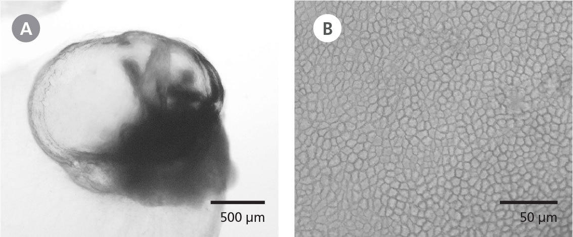
図2. 脈絡叢オルガノイドに見られる液体で満たされた嚢胞と上衣上皮の形態
(A) STEMdiff™ Choroid Plexus Organoid DifferentiationおよびMaturation Kitで分化・成熟させた培養40日目の脈絡叢オルガノイド全体の位相差画像。脳脊髄液(CSF)に似た液体を内包する半透明の嚢胞が確認できる。(B) 脈絡叢オルガノイドの上皮は、生体内で脳室を覆う上衣細胞に典型的な、密に詰まった立方状形態を示す。
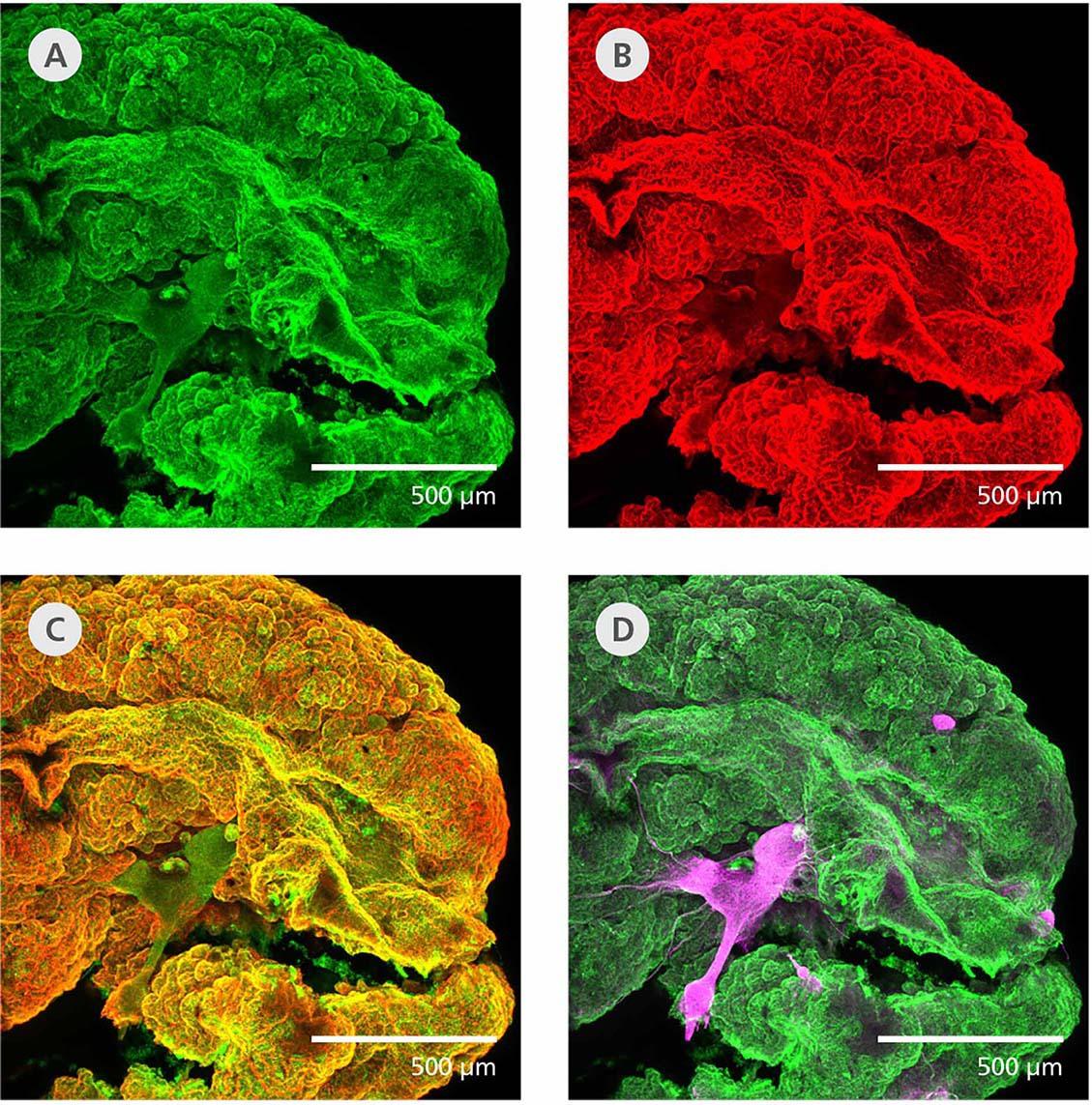
図3. 脈絡叢オルガノイドの上衣上皮マーカー発現と、限定された神経上皮マーカー発現
培養40日目の脈絡叢オルガノイド全体を透明化・染色し(Masselink et al. 2019)、免疫組織化学的に解析した。嚢胞構造は(A)TTR(緑)および(B)CLIC6(赤)陽性であった。(C) これらの上衣細胞マーカーは高度に共局在している。(D) MAP2陽性(マゼンタ)のニューロンの束が見られるが、上衣細胞マーカーとは共局在していない。
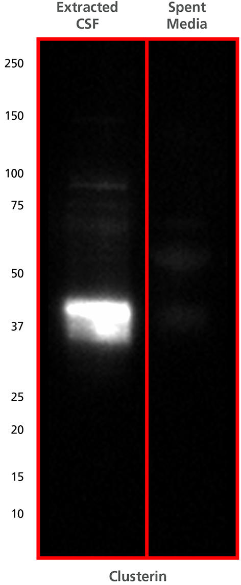
図4. 脈絡叢オルガノイドの嚢胞からの抽出液には、CSFマーカーであるClusterinタンパク質が豊富に含まれる
培養40日目の脈絡叢オルガノイドの嚢胞から、脳脊髄液(CSF)様の液体を28Gシリンジで抽出した。抽出液を用いてClusterinを検出するウエスタンブロットを行うと、分子量マーカー37~50 kDaの間にバンド1本が確認された。Clusterinは、CSF中に高濃度で存在する可溶性の分泌シャペロンタンパク質である。
本品のベースとなる組成を開発したPellegrini博士のウェビナー:
Modeling Brain Barrier Permeability and CSF Secretion with Choroid Plexus Organoids
製品記事
神経研究に有用な培養ツールまとめ(STEMCELL Technologies社)
オルガノイド研究向けの培養ツールまとめ(STEMCELL Technologies社)
ES/iPS細胞からさまざまな細胞への分化誘導(STEMCELL Technologies社)
ヒトES/iPS細胞における品質管理の考え方:細胞品質管理のタイミング
「mTeSR Plus-cGMP」:週2回の培地交換で高品質なES/iPS細胞を!
「mTeSR1」フィーダー細胞が不要なヒトES/iPS細胞維持用 無血清培地