STEMCELL Technologies STEMdiff Human iPSC-Derived Astrocytes
- 研究用
- 新製品
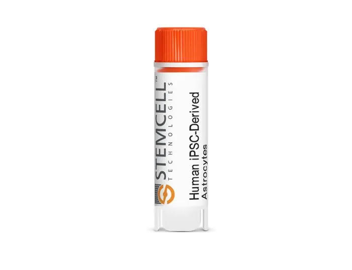
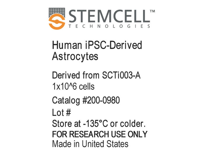
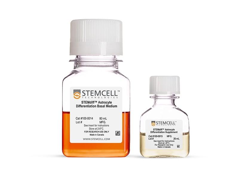
Human iPSC-Derived Astrocytesは、ただちに使用可能な高品質・高純度(S100B陽性率≥70%、GFAP陽性率≥60%、DCX陽性ニューロン前駆細胞率<19%)の凍結保存ヒトグリア細胞です。本製品は詳細に特性評価されたヒトiPSCコントロール株SCTi003-Aより、STEMdiff™ SMADi Neural Induction Kit、 STEMdiff™ Astrocyte Differentiation Kit、およびSTEMdiff™ Astrocyte Serum-Free Maturation Kitよりなる血清フリーのシステムで製造されています。STEMdiff™ Astrocyte Serum-Free Maturation Kitを使用して起眠後、直ちに下流アプリケーションに利用可能です。ヒト神経発達・疾患のモデル化、共培養応用、薬剤スクリーニング、毒性試験、細胞治療の検証など、多様なツールとして活用いただけます。
本製品は研究用製品(RUO)であり、学術・商業研究用としての承認を受けています。血液サンプルは、治験審査委員会(IRB)またはその他の規制当局が承認した同意書およびプロトコルを用いて倫理的に提供されています。
製品の特長
ヒトiPS細胞株 SCTi003-Aから分化したアストロサイト
- すぐに使える凍結保存ヒトアストロサイトで時間を節約
- 完全に無血清のシステムで製造されたアストロサイトの高純度と機能性を実現
- STEMdiff™ Astrocyte Serum-Free Maturation Kitでヒトアストロサイトの長期培養・維持可能
- 同一の遺伝的背景をもつ適切な神経細胞-アストロサイトの共培養系樹立が可能
- 神経発生、疾患、薬物スクリーニング、毒性、細胞治療のアプリケーションのモデル化に柔軟に対応
ご注意
ソースセルバンクのドナー詳細および細胞品質特性については、以下のSCTi003-Aの情報をご参照ください。
コントロールに最適な凍結ヒトiPS細胞株
SCTi003-Aは、αβT細胞に由来し、VDJ配列の再構成を受けています。核型的に安定であり、3胚葉分化能を示し、未分化細胞マーカーを発現し、非組み込み型技術によりリプログラミングされています。hPSCreg®に登録されており、コミュニティ基準に基づく倫理的・生物学的適合性が保証されます。詳細については、ロット別の検査証明書(CoA)およびiPSC株に関するよくある質問もご参照ください。
データ紹介
Figure 1. Human iPSC-Derived Astrocytes Exhibit High-Quality Morphology
Cryopreserved Human iPSC-Derived Astrocytes generated from SCTi003-A iPSCs were thawed and plated onto Matrigel®-coated plates at 150,000 cells/cm² in STEMdiff™ Astrocyte Serum-Free Maturation Kit. Astrocytes were incubated at 37°C and subsequently analyzed on Days 1, 3, and 7 by brightfield microscopy at 10x magnification. Astrocytes display the expected morphology, including a flattened, polygonal shape with enlarged cell bodies, and by Day 7, more pronounced stellate processes characteristic of mature astrocytes. iPSC = induced pluripotent stem cell.
Figure 2. Human iPSC-Derived Astrocytes Exhibit High Purity and Express Characteristic Glial Markers
Cryopreserved Human iPSC-Derived Astrocytes generated from SCTi003-A iPSCs were thawed and cultured using the STEMdiff™ Astrocyte Serum-Free Maturation Kit for 7 days and fixed for immunocytochemistry. (A) Day 7 astrocytes show high expression of astrocyte markers S100B (green) and GFAP (red), with low expression of the neuronal marker DCX (purple) (20× magnification, scale bar = 500 µm). (B) The percentage of cells positive for S100β, GFAP, and DCX was quantified from five images per condition across three independent experiments, expressed relative to total DAPI-positive cells. S100β was expressed in 84% of cells, GFAP in 79%, and DCX in fewer than 3%. Error bars represent standard deviation (n = 3 biological replicates). iPSC = induced pluripotent stem cell.
Figure 3. Human iPSC-Derived Astrocytes Express Expected Levels of Genes Characteristic for Astrocytes
Cryopreserved Human iPSC-Derived Astrocytes generated from SCTi003-A iPSCs were thawed and cultured using the STEMdiff™ Astrocyte Serum-Free Maturation Kit for 7 days prior to analysis. Expression levels were measured by quantitative PCR (qPCR) and normalized to hPSC controls relative to housekeeping gene TBP. iPSC = induced pluripotent stem cell; hPSC = human pluripotent stem cell.
Figure 4. Human iPSC-Derived Astrocytes Exhibit High-Quality Morphology
Cryopreserved Human iPSC-Derived Astrocytes generated from the SCTi003-A iPSCs were thawed and cultured in STEMdiff™ Astrocyte Serum-Free Maturation Kit for 7 days. Prior to glutamate treatment, cells were incubated in BrainPhys™ medium for 4 hours at 37°C and 5% CO₂. Cells were then treated for 1 hour with either 50 µM glutamate in BrainPhys™ ("GLU") or water in BrainPhys™ ("CTRL"). Glutamate levels in each sample were quantified using the Fluorometric Glutamate Assay Kit (Abcam, ab138883). Different colors represent distinct experimental setups. Bars represent mean ± standard deviation. (A) Glutamate concentration in the media (“Pre-Treatment”) and spent media (“Post-Treatment”). (B) Glutamate concentration in the cell lysates, extracted with Mammalian Cell Lysis Buffer (ABCAM, ab179835). iPSC = induced pluripotent stem cell.
Figure 5. Human iPSC-Derived Astrocytes Display Reactive Phenotype Upon Cytokine Stimulation
Cryopreserved Human iPSC-Derived Astrocytes generated from the SCTi003-A iPSCs were thawed and cultured in STEMdiff™ Astrocyte Serum-Free Maturation Kit for 7 days. Astrocytes were treated with either a vehicle control ("CTRL") or a pro-inflammatory cytokine cocktail ("A1") consisting of 30 ng/mL TNFα, 3 ng/mL IL-1α, and 400 ng/mL C1Qa for 24 hours. Following A1 treatment, astrocytes showed increased expression of C3 (marker of neurotoxic reactive astrocytes) and GBP2 (interferon response and inflammation marker). Expression levels were measured by quantitative PCR (qPCR) and normalized to hPSC controls relative to housekeeping gene TBP. Statistical significance was determined using an unpaired t-test (***p < 0.001). Bars represent mean ± standard deviation, with different colors indicating distinct experimental conditions. iPSC = induced pluripotent stem cell.
Figure 6. Human iPSC-Derived Astrocytes Can Be Co-Cultured with Neurons to Model Cell-Cell Interactions In Vitro
Representative immunocytochemistry images display Human iPSC-Derived Astrocytes co-cultured with forebrain neurons derived using the STEMdiff™-TF Forebrain Induced Neuron Differentiation Kit for 21 days and maintained in the STEMdiff™ Forebrain Neuron Maturation Kit. Cells were stained for GFAP (green, astrocyte marker) and MAP2 (pink, neuronal dendrite marker). The presence of MAP2-positive neurons alongside GFAP-positive astrocytes demonstrates compatibility in co-culture conditions, supporting the viability and integration of neurons within an astrocyte-rich environment. Scale bar = 100 µm.
Figure 7. Human iPSC-Derived Astrocytes Can Be Tri-Cultured with Neurons and Microglia to Model Cell-Cell Interactions In Vitro
Representative immunocytochemistry images display Human iPSC-Derived Astrocytes tri-cultured with forebrain neurons derived using the STEMdiff™-TF Forebrain Induced Neuron Differentiation Kit and microglia for 7 days using BrainPhys™ hPSC Neuron Kit supplemented with STEMdiff™ Microglia Supplement 2 (Component #100-0023 of STEMdiff™ Microglia Differentiation Kit). Cells were stained for GFAP (green, astrocyte marker), MAP2 (pink, neuronal dendrite marker), IBA1 (cyan, common microglia marker), and DAPI (grey, nuclei). The presence of MAP2-positive neurons, GFAP-positive astrocytes, and IBA1-positive microglia confirms the successful establishment of tri-culture conditions and supports the viability and integration of astrocytes in a mixed neuron–microglia environment. Scale bar = 50 µm.
Figure 8. Human iPSC-Derived Astrocytes Promote Neural Activity in Co-Culture with Human iPSC-Derived Neurons
Human iPSC-Derived Forebrain Neuron Precursor Cells (Catalog #200-0770) were cultured either alone or in a 1:1 co-culture with isogenic Human iPSC-Derived Astrocytes (Catalog #200-0980). Cells were plated on a 48-well CytoView MEA™ Plate (Catalog #200-0872) and maintained in STEMdiff™ Forebrain Neuron Maturation Medium (Catalog #08605). Electrical activity from 16 electrodes per well was recorded longitudinally using the Maestro Pro™ MEA system (Catalog #200-0887). (A - B) Representative brightfield images show the morphology of neuron-only and neuron-astrocyte co-cultures on the MEA plate. (C - D) Raster plots on Day 21 illustrate spike activity in neuron-only versus co-culture conditions. Detected spikes (black lines), single-channel bursts (blue lines; defined as ≥5 spikes with inter-spike intervals [ISI] ≤100 ms), and network bursts (≥50 spikes across ≥35% of electrodes with ISI ≤100 ms) were quantified over time. Co-cultures showed a visibly higher level of neuronal activity on Day 21 compared to neurons alone. (E - G) Quantitative analyses revealed a progressive increase in (E) mean firing rate, (F) number of active electrodes, and (G) burst frequency over the 21-day period. Across all measures, neuron-astrocyte co-cultures consistently exhibited significantly greater electrophysiological activity than neuron-only cultures. hiPSC = human induced pluripotent stem cell; MEA = microelectrode array; ISI = inter-spike interval.

