STEMCELL Technologies BrainPhys BrainPhys Imaging Optimized Medium
- 研究用
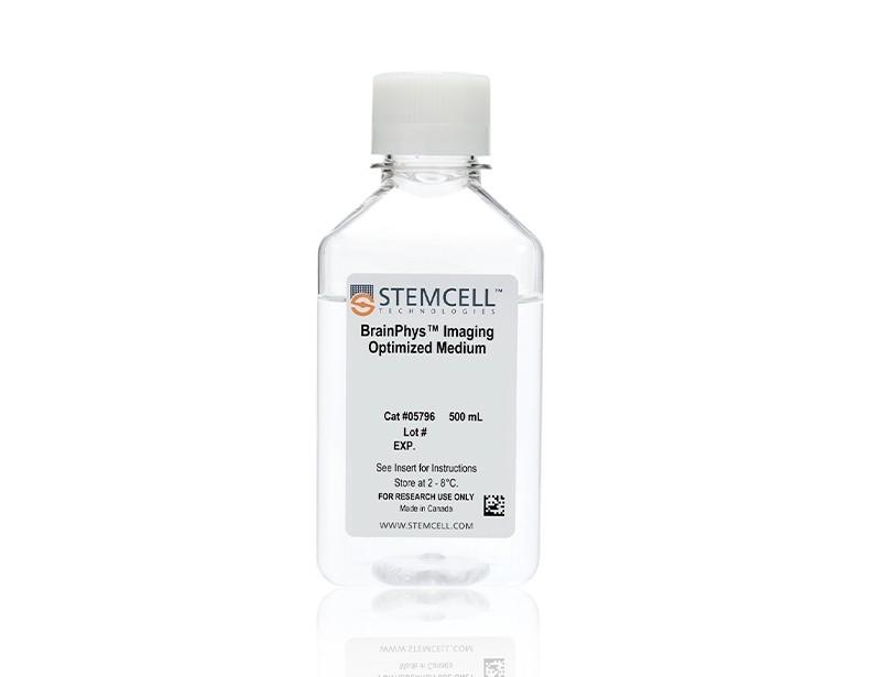
BrainPhys™ Imaging Optimized Medium(ST-05796)は、神経細胞用の無血清基礎培地です。Cedric BardyとFred H. Gageが報告したBrainPhys™に基づく組成で、イメージング用途向けに最適化されています。
本品のアプリケーションには、ライブ蛍光イメージング(カルシウムイメージングおよび光遺伝学)や神経細胞培養が含まれます。培地組成からフェノールレッドを除去したことに加え、バックグラウンドの蛍光を低減し、連続露光時の安定性を高めるように改変されています (M Zabolocki et al. Nat Comm, 2020)。
製品の特長
BrainPhys™ Imaging Optimized Mediumで、ニューロンのライブイメージングと神経機能を改善
- 長時間のライブイメージングで光毒性なし
- 488 nm励起チャネルにおける自家蛍光バックグラウンドを低減
- 培養と同条件下で機能アッセイを行えるため、培地交換による細胞への負荷を回避
- 脳の細胞外環境をより適切に再現
- 神経機能の改善、およびシナプス活性の高いニューロン比率の増加
- 厳密な原料スクリーニングと品質管理で、製品ロット間差を最小化
生細胞イメージングの流れ
hPSC由来ニューロンの分化過程でのライブイメージング
hPSC由来ニューロンを、適切なサプリメントを加えたBrainPhys Imaging Optimized Mediumに移行して最大14日間培養できます。
プライマリー組織由来ニューロンの成熟過程でのライブイメージング
プライマリー組織由来ニューロンを、血清代替サプリメントを加えたBrainPhys Imaging Optimized Mediumに移行して最大14日間培養できます。
データ紹介
イメージングする細胞の光毒性と自家蛍光を低減
(A)BrainPhys Imaging Optimized Mediumで培養した初代ラット皮質ニューロンは、青色LED光に12時間露光した後も健康な形態を維持します。(B)生きたニューロンを染色するNeuroFluor™ NeuO色素で標識したニューロンは、平均発光波長525 nmのバックグラウンド蛍光が減少し、画像のコントラストが改善しました。元のBrainPhys Neuronal Mediumよりもライブイメージング条件下で優れた性能を示しています。
元のBrainPhys Neuronal Mediumは長期培養で細胞を健康に保ちますが、同条件で(C)崩壊した細胞体と神経突起(黒矢印)、および(D)自家蛍光を示します。
標準的な神経培地や他社製イメージング培地より自家蛍光を低減
The emission spectra across 400 - 700 nm of the light spectrum were captured as mean autofluorescence intensity from test and control (PBS) media (without cells) for the 375 (ultraviolet), 405 (violet), 488 (blue), and 532 nm (green) excitation wavelengths. Mean fluorescence intensity in PBS was subtracted from other media to normalize the data (n = 8 replicate wells, 3 independent experiments). BrainPhys™ Imaging Optimized (BPI) shows autofluorescence intensities similar to PBS and far lower intensities than are observed from standard neural media. Adapted from Zabolocki et al. 2020, Nature Communications, available under a Creative Commons 4.0 License.
3D神経培養の蛍光イメージングで、シグナル対バックグラウンド比を改善
GFP-labeled ventral forebrain organoids were co-cultured and fused with unlabeled dorsal forebrain organoids for one week prior to live imaging in Forebrain Organoid Differentiation Medium from STEMdiff™ Dorsal Forebrain Organoid Differentiation Kit (right) or BrainPhys™ Imaging Optimized Medium (BPI, left). Interneuron migration can be visualized more clearly in BPI. Adapted from Zabolocki et al. 2020, Nature Communications, available under a Creative Commons 4.0 License.
ヒトニューロンのin vitro ライブカルシウムイメージング
Intracellular Ca2+ changes in human PSC-derived neurons were measured with time-lapse imaging sequences of a Ca2+ sensor. Regions of interest (ROIs) were drawn on the cell soma to determine fluorescence intensity changes (ΔF/F0) over time. The same fields of view (FOVs) were imaged in artificial cerebrospinal fluid (ACSF), BrainPhys™ Imaging Optimized (BPI), Imaging Alternative 2, and Imaging Alternative 1 media. (A) Representative image of a neuronal population in BPI. White circles represent active ROIs, and fluorescent intensity represents intracellular Ca2+ levels. (B) Comparison of Ca2+ signals in BPI, Imaging Alternative 1 (Img Alt 1), and Imaging Alternative 2 (Img Alt 2) conditions show significant differences in the proportions of cells with Ca2+ spikes (two-tailed Mann-Whitney tests, mean ± SEM). Active cells with Ca2+ spikes in BPI (n = 243 cells), Imaging Alternative 2 (n = 39 cells), and Imaging Alternative 1 (n = 18 cells) were compared across seven FOVs from two coverslips. (C) Example traces from the same ROIs in different media. Adapted from Zabolocki et al. 2020, Nature Communications, available under a Creative Commons 4.0 License.
神経活動のイメージングに基づく長期解析
Spontaneous firing rates of human neurons expressing a synapsin:ChETA-YFP optogene were recorded in BrainPhys™ (BP), BrainPhys™ Imaging Optimized (BPI), or Imaging Alternative 1 media with identical supplements using microelectrode arrays. Recordings were split into two groups (circles, n = 8 wells; or triangles, n = 7 wells). Both groups were switched from BP to BPI after 82 days, and only the triangle group was switched to Imaging Alternative 1 from day 89 - 95. From day 96, both groups were cultured in BP for one week before switching to BPI. Long-term firing is enhanced in BPI relative to Imaging Alternative 1, and mean firing rates (MFR, mean ± SEM) from both groups perform at baseline following the media change at day 96. Adapted from Zabolocki et al. 2020, Nature Communications, available under a Creative Commons 4.0 License.
ヒト神経機能のin vitro 長期解析(BrainPhys™ Neuronal Mediumと同等性能)
Multielectrode array (MEA) recordings from human pluripotent stem cell (hPSC)-derived neurons cultured in a 48-well MEA plate in BrainPhys™ (BP) for 9 weeks (n = 6 wells, 96 electrodes) or switched to BrainPhys™ Imaging Optimized (BPI) from BP during weeks 6 - 8 (n = 5 wells, 80 electrodes). (A) Percentage of active electrodes (>0.017Hz) and (B) mean firing rate (MFR) were both maintained during the period in BPI. Values are presented as mean ± SEM. Adapted from Zabolocki et al. 2020, Nature Communications, available under a Creative Commons 4.0 License.

